X-rays. Рентген-снимки
1) В советско-снг-вской ортопедии (и не только ортопедии) информация пиздилась из-за рубежа без указания источников ВСЕГДА. Все книги по ортопедии писались и продолжают писаться как застольные тосты - несётся всякая ахинея. Изредка действительно даются ссылки на некие исследования, но в основном даваемая информация - беспочвенное безграмотное пиздоболство. Пример тому - начиная от "классики советской ортопедии" В.О. Маркса, 1978 и заканчивая свежаком - «Травматология и ортопедия». Редакторы: чл.-корр РАМН, засл. деят. науки РФ профессор Н.В. Корнилов и профессор Э.Г. Грязнухин, Спб, изд. «Гиппократ», 2006 с их предложениями решать все проблемы со сколиозом с помощью корсета Шено и совершенно необоснованно сообщая о его эффективности.
2) Отсутствие возможности изучать языки, в частности, английский, и недоступность зарубежных источников информации позволяла этому мошенничеству сходить врачам-пейсателям в рук десятилетями.
3) Всё это способствовало тому, что задержка между получениями результатов исследований за рубежом и их применению на практике достигала 50 (прописью - пятидесяти) лет.
4) Сейчас я ОЧЕНЬ часто сталкиваюсь с назначением в диагностических целях снимка лёжа, причём, иногда без сопутствующего снимка стоя.
Пятьдесят, если не шестьдесят лет назад, в странах, где проводятся исследования, такой метод диагностики действительно использовался. Лет 50 назад. Но им с тех пор никто не пользуется, потому что он НЕинформативен, а значит - это просто лишняя лучевая нагрузка на организм.
Тупость назначающих и непонимание что, как и для чего делается не перестают меня потрясать. Начиная с того, что снимки делают "по отделам" ("а чё такова, у нас плёнка короткая, как мы снимем весь позвоночник" - руками, бля. Возьмёте и закрепите две подряд кассеты руками) вплоть до того, что ОДИН отдел снимают лёжа, ВТОРОЙ - стоя, и потом по этим снимкам ставят диагноз!!!
Я не говорю о такой мелочи, как вечное гадание где правая сторона, а где левая - это же так трудно - завести хотя бы одну метку на рентген-кабинет и всегда ставить её в последний момент! Пришёл снимок - метка изначально была на плёнке. Перепутала стороны клуша или нет при заправлении и установке кассеты - теперь загадка.
Обычно право-лево определяют по тени желудка, он полый и более тёмный, чем печень справа. Желудок расположен слева. Но эти тени не всегда очевидны.
Выполнение рентгена спины:
Ситуация такова:
Для определения возможности коррекции дуги смотрят на её подвижность. Главным образом, смысл смотреть на подвижность дуги присутствует перед операцией. Чем более подвижна дуга - тем лучше будет коррекция в результате операции.
При корсетировании и прочих консервативных методах лечения глубокого смысла в рассматривании подвижности дуги нет, т.к. большая её подвижность означает также и бОльшую вероятность прогрессии по сравнению с фиксированной (структурной) дугой.
Определение подвижности может быть полезным при S-сколиозе когда обе дуги симметричны. При этом более ригидная (неподвижная) дуга является более ранней и, скореё всего, первичной.
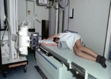
ФОТО: http://www0.hku.hk/ortho/spine.html
Fulcrum bending radiograph for assessing spinal flexibility in scoliosis (Fulcrum (англ.) - точка опоры)
Так вот, как определяют подвижность позвоночника:
1) Делают снимок стоя
2) Делают спец. снимок - снимок с максимально достижимой корекцией
3) Сравнивают их. Если форма дуги меняется - она подвижна. Если не меняется - неподвижна, структурная. Ну, и соответственно, бывает разная степень подвижности.
Виды спецснимков:
1) Стоя в наклоне
2) Side bending - лёжа с наклоном
3) Fulcrum bend - лёжа с подкладыванием подушечек и валиков
4) Push prone - лёжа на животе, кто-то давит на самую выпуклость, корректируя таком образом ротацию.
5) UGA traction - максимальная вытяжка под анестезией.
Результаты разных исследований по сравнению этих методов между собой:
Исследования эффективности снимка "стоя в наклоне" я не увидел вообще. Скорей всего, не применяется широко, т.к. даёт не максимальную коррекцию
1) Четвёртый тип (Push prone) и второй методы (Side bending - лёжа с наклоном) дают одинаковую коррекцию (см. по порядку ссылки ниже, начиная с Пабмедовских).
2) Эффективность третьего (Fulcrum bend - лёжа с подкладыванием подушечек и валиков) выше, чем второго (Side bending - лёжа с наклоном), а эффективность второго выше, чем пятого (UGA traction - максимальная вытяжка под анестезией).
3) Пятый (UGA traction - максимальная вытяжка под анестезией) по эффективности равен второму (Side bending - лёжа с наклоном) для грудных и грудо-поясничных дуг и эффективность изменения сравнима с четвёртым методом (Push prone - лёжа на животе, кто-то давит на самую выпуклость, корректируя таком образом ротацию) для неструктурных грудо-поясничных дуг.
4) Четвёртый тип (Push prone) даёт большую коррекцию, чем второй метод (Side bending - лёжа с наклоном)
5) Третий тип (Fulcrum bend - лёжа с подкладыванием подушечек и валиков) лучше работает при грудных дугах среднего размера, чем второй (Side bending - лёжа с наклоном). Эти два метода имеют одинаковую эффективность в коррекции поясничных дуг.
6) Исключительно чтобы не заниматься cherry-picking:
http://www.ncbi.nlm.nih.gov/pubmed/18007242
CONCLUSION:
A single preoperative supine radiograph is highly predictive of side-bending radiographs and can be used as an adjunct to predicting curve type, flexibility, and structurality. Thus, this singular, reproducible, and non-effort-related radiograph can potentially replace the need for dual side-bending films.
Корейцы провели исследование и пришли к выводу, что одного снимка лёжа будет достаточным для определния подвижности дуги. Однако, странность методологии исследования не позволяет мне ему доверять.
Итого: для определения подвижности дуги просто снимок лёжа используется только в СНГ и может быть, в Корее. Специалисты более развитых стран используют более продвинутые методы выполнения снимков. Снимок лёжа либо имеет смысл делать с максимально возможной коррекцией, либо не имеет смысл делать вообще. Наибольшее распространение получил метод выполнения снимка лёжа с наклоном ввиду сочетания простоты, повторяемости и эффективности.
http://books.google.com/books?id=cZu3_EezS_wC&pg=PA79&lpg=PA79&dq=Push-prone+radiographs&source=bl&ots=JsdJvoyh7T&sig=mvRWHZISuC__9J9kmwhlt4yIZlM&hl=en&ei=uh43Tr2lB4rliAKa7LT4Dg&sa=X&oi=book_result&ct=result&resnum=6&sqi=2&ved=0CEkQ6AEwBQ#v=onepage&q=Push-prone%20radiographs&f=false
Spinal deformities: the essentials
By Robert F. Heary, Todd J. Albert
Radiologic Evaluation of the Spinal Deformity: Is It Fixed or Flexible?
The rigidity or the flexibility of the spinal deformity should be evaluated in all curves that are considered for surgical intervention. Certain techniques of radiographic evaluation have advantages over others depending on the curve characteristics and patient compliance. Traction films may be suitable for cooperative pediatric patients with large and prominent coronal curve or proximal thoracic curves.
The use of a bolster placed under the apex of the deformity to maximize postural correction is particularly useful in cases of kyphotic deformity in adolescent or adult patients. This technique permits a better assessment of curve flexibility than can be performed by the patient attempting correction by maximally extending the spine. Literature on radiographic assessment of curve flexibility principally discusses methods of evaluation used in assessing adolescent idiopathic scoliosis and neuromuscular scoliosis. There is unfortunately little data critically comparing the effectiveness of radiographic methods of determining curve flexibility in the adult spinal deformity patient.
In all cases, the initial assessment involves upright full-length 36-in. x 14-in. scoliosis films with both posterior/anterior (PA) and lateral views. Because curve magnitudes may vary with the elimination of gravity in supine radiographs, these should be considered in adult scoliosis patients. To optimize visualization for the lateral radiograph, "clavicle position," where the patient fully flexes the elbows with the hands in a relaxed fist, wrists flexed, placing the proximal interpha- langeal (PIP) joints comfortably up into the supraclavicular fossa, thus passively flexing the humerus forward, produces significantly better overall visualization of critical vertebral landmarks compared with positions where the arm is either straight out or partially flexed.(16)
Ideally, the lateral radiograph should visualize C2 to the pelvis including the femoral heads to assess the sagittal balance of the spinal column. Similarly, on the PA view, the margins of the rib cage and the pelvis along with the femoral heads should be clearly visualized. This will facilitate the assessment of the skeletal maturity of a pediatric patient. The visualization of the ribs helps to determine the associated thoracic cage deformity. Both a congenital fusion of the ribs or significant chest wall deformity are associated with rigid or fused spinal segments. After assessing the spinal balance on the sagittal and coronal planes, the Cobb measurements are obtained for each area of the spine (cervical, proximal thoracic, main thoracic, and thoracolumbar/lumbar). The amount of vertebral body rotation at the apex of the coronal curve also determines the rigidity of the curve. The greater the vertebral body rotation, the greater the rigidity of the coronal curve. If the curve and clinical characteristics warrants the possibility of requiring surgical intervention, then the flexibility of the curve should be performed.
Flexibility of the coronal plane deformity is most commonly measured by supine PA left and right lateral bending films. The supine position is recommended so the patient can give a maximal effort without the possibility of falling. This position also eliminates the effect of gravity and will further clarify the flexibility of the deformity. Optimally, the full-length scoliosis films should be used to permit assessment of the flexibility of all areas of the deformity. Traction and push-prone radiographs have been used by some deformity surgeons to measure the flexibility of the thoracic and lumbar curves.(24-25) Traction films are also used to determine the flexibility of the primary and compensatory curves and to assess the inherent capability of the curve to correct with traditional spinal instrumentation techniques. This enables the spine surgeon to determine the extent of intraoperative correction maneuvers that will be necessary to achieve spinal balance both in the sagittal and coronal planes. Traction radiographs are performed by the surgeon pulling on the head and neck while countertraction is applied to the lower extremities (Figs. 9-3A to 9-3E).(26)
A push-prone radiograph is performed with patient in a supine position with manual force placed over the apex of the spinal deformity. This radiographic technique can determine both the primary curve flexibility and the compensatory curve response with forceful correction of the primary curve.(25) For this particular curves pattern in a patient with AIS the push-prone radiograph most accurately predicts the position of the last instrumented vertebrae when treated with anterior spinal fusion and instrumentation. The technique involves translational forces applied at the apex with counterstabilizing forces applied at the axilla and pelvis on the opposite side.
Fulcrum-bending films are similar to bolster films used for kyphosis correction and are advocated by some authors in assessing scoliosis. This method involves placing the patient in the lateral decubitus position and bending over a fulcrum (a radiolucent padded cylinder) so that the spine is passively hinged open. The fulcrum-bending radiograph has been shown to be more predictive of the degree of flexibility and correctability than the lateral-bending radiograph in this group of patients who had segmental spinal instrumentation for correction of idiopathic scoliosis.(27) These films are thought to be more effective in predicting curve responses to surgical intervention. Halo gravity traction is an option for assessing very large curves and rigid curves particularly in patients with severe scoliosis, kyphoscoliosis, and kyphosis. It has been found to be a safe, well-tolerated method of applying gradual, sustained traction to maximize preoperative evaluation and postoperative correction in this difficult population.(28)
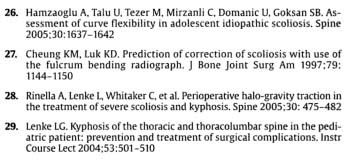
26. Hamzaoglu A. Talu U. Tezer M. Mirzanli C. Domanic U. Goksan SB. Assessment of curvc flexibility in adolescent idiopathic scoliosis. Spine 2005:30:1637-1542
27. Cheung KM. Luk КГ). Prediction of correction of scoliosis with use of the fulcrum bending radiograph. J Bone Joint Surg Am 1997:79: 1144-1150
28. Rinella A. tenke L, Whilaker С. et al. Perioperative halo-gravity traction in the treatment of severe scoliosis and kyphosis. Spine 2005:30: 475-482
29. Lenke LC. Kyphosis of the thoracic and thoracolumbar spine in the pediatric patient: prevention and treatment of surgical complications. Instr Course lect 2004:53:501-510
http://www.knowyourback.org/Pages/SpinalConditions/Scoliosis/default.aspx (The clinical information provided on this website is developed and reviewed by the physician members and spine specialists of the North American Spine Society, a nonprofit, multidisciplinary organization dedicated to spine education, research and advocacy.)
While the patient is standing, X-ray studies are taken with the patient standing in
- the anterior/posterior plane,
- the lateral plane,
- side bending,
- fulcrum bend and
- push prone. During the push prone X-ray study, the patient is placed on the radiograph table in the prone (on their belly) position while manual pressure is applied to the apex of the thoracic curve at the same time the pelvis and shoulders are stabilized.
The side bending X-ray studies help to determine the flexibility of the curves.
http://www.ncbi.nlm.nih.gov/pubmed/10647164
CONCLUSIONS:
The push-prone and lateral-bending radiographs are similar in predicting less correction of the Cobb angle after anterior spinal surgery.
http://www.ncbi.nlm.nih.gov/pubmed/2336909
The authors evaluated a group of 297 patients after the surgical correction and stabilization of the spine, out of which there were 171 idiopathic, 98 congenital, 14 neuromuscular deformities and 14 deformities were connected with neurofibromatosis. By the statistical comparison of the values of curvature when the patient is standing prior to operation, lying in distraction of 200N and lying in side-bend to the convex of the curve with the value of correction after operation the authors have proved that it is sufficient to carry out only the examination in the maximum voluntary side-bend with the exception of the deformities of neuromuscular origin where it is necessary to perform also x-ray examination in the distraction of 200N when the patient is lying.
http://www.ncbi.nlm.nih.gov/pubmed/16025034
METHODS:
A total of 34 consecutive patients with AIS who had surgical treatment were studied. Preoperative radiologic evaluation consisted of
standing
- anteroposterior and
- lateral,
supine
- lateral bending and
- traction,
- fulcrum bending radiographs, and also
- supine traction radiographs taken with the patient under general anesthesia (UGA) just before surgery. All structural curves were measured, and the flexibility ratio was determined on each radiograph.
CONCLUSION:
Fulcrum higher than bending higher than traction with the patient UGA is the order of radiographs for better predicting flexibility and correction in curves between 40 degrees and 65 degrees. Flexibility obtained at traction radiographs with the patient UGA is clearly better in numerical values, and closer to the amount of surgical correction than the amount of flexibility at fulcrum and side-bending radiographs for curves larger than 65 degrees, although not statistically significant as a result of the small number of patients in this group.
http://www.ncbi.nlm.nih.gov/pubmed/16477449
The pre-operative
- standing postero-anterior and lateral radiographs,
- supine side bending radiographs,
http://www.ncbi.nlm.nih.gov/pubmed/20110844
RESULTS:
Traction UGA demonstrated equal flexibility to supine bending in structural MT and TL/L curves, with a trend towards more flexibility in severe MT curves > or = 60 degrees, while push-prone demonstrated significantly less flexibility. Each of the methods showed significantly less flexibility than postoperative correction in MT curves, while traction UGA and supine bending were not significantly different than postoperative correction in TL/L curves. Analysis of nonstructural TL/L curves showed that traction UGA and push-prone were not significantly different than postoperative correction, while supine bending overestimated postoperative correction.
CONCLUSION:
Traction UGA offers flexibility equivalent to supine bending for structural MT and TL/L curves, and flexibility comparable with push-prone for nonstructural TL/L curves. Traction UGA also shows both structural and compensatory curves on the same radiograph, and ultimately may provide a better estimate of spinal balance.
http://www.ncbi.nlm.nih.gov/pubmed/17173001
SUMMARY OF BACKGROUND DATA:
Repeated radiographic examinations at young age may increase the risk for breast cancer in adulthood. MRI images the spine satisfactorily but is cumbersome in standing. A harness supplying axial load to a lying subject simulates the standing radiograph appearance of the lumbar spine.
METHODS:
Thirty patients with idiopathic scoliosis greater than 20 degrees performed a routine posteroanterior and lateral standing thoracolumbar spine radiograph and an MRI in supine position without and with axial loading.
RESULTS:
Mean Cobb angle for the major curve was 31 degrees on standing radiographs, 23 degrees on nonloaded supine MRI, and 31 degrees on supine loaded MRI. Axially loaded, compared with nonloaded, MRI increased the Cobb angle by 8 degrees. The mean difference between standing radiograph and supine axially loaded MRI was 0 degrees, with an intermethodologic variation(s) of 3.4 degrees. Radiographic and MRI (axially loaded) Cobb angles correlated positively (r = 0.78).
CONCLUSIONS:
Axial loading on supine MRI produces coronal Cobb angles similar to standing radiographs. This is a way to acquire reliable Cobb angles without radiation in the monitoring of idiopathic scoliosis.
http://journals.lww.com/spinejournal/Abstract/2000/01010/Comparison_of_Push_Prone_and_Lateral_Bending.14.aspx
Results. The lateral-bending and the push-prone radiographs predicted less correction of the Cobb angle and the angle of the lowest instrumented vertebra to the horizontal than was achieved after surgery. However, the push-prone radiograph was superior to the lateral-bending radiograph in accurately predicting the postoperative correction of the rotation of the lowest instrumented vertebra as well as the translation of the lowest instrumented vertebra from the center sacral line.
Conclusions. The push-prone and lateral-bending radiographs are similar in predicting less correction of the Cobb angle after anterior spinal surgery. The push-prone radiograph helps in determining the effects that correction of the primary curve has on the curves above and below the level of fusion by better predicting the translational correction of the lowest instrumented vertebra and the rotation of the lowest instrumented vertebra.
http://www.ncbi.nlm.nih.gov/pubmed/18067811
OBJECTIVE:
To compare the effects in assessing the curve flexibility of the adolescent idiopathic scoliosis (AIS) and predicting the outcomes of operation among different radiological techniques: supine lateral bending (SB), traction (Tr), and fulcrum bending radiographs.
CONCLUSION:
Fulcrum-bending radiography can better assess the flexibility and correction rate of thoracic curves in AIS, however, it can only predict those in moderate thoracic curves. Fulcrum-bending radiograph and SB radiograph are similar in predicting the flexibility in lumbar curves.
2) Отсутствие возможности изучать языки, в частности, английский, и недоступность зарубежных источников информации позволяла этому мошенничеству сходить врачам-пейсателям в рук десятилетями.
3) Всё это способствовало тому, что задержка между получениями результатов исследований за рубежом и их применению на практике достигала 50 (прописью - пятидесяти) лет.
4) Сейчас я ОЧЕНЬ часто сталкиваюсь с назначением в диагностических целях снимка лёжа, причём, иногда без сопутствующего снимка стоя.
Пятьдесят, если не шестьдесят лет назад, в странах, где проводятся исследования, такой метод диагностики действительно использовался. Лет 50 назад. Но им с тех пор никто не пользуется, потому что он НЕинформативен, а значит - это просто лишняя лучевая нагрузка на организм.
Тупость назначающих и непонимание что, как и для чего делается не перестают меня потрясать. Начиная с того, что снимки делают "по отделам" ("а чё такова, у нас плёнка короткая, как мы снимем весь позвоночник" - руками, бля. Возьмёте и закрепите две подряд кассеты руками) вплоть до того, что ОДИН отдел снимают лёжа, ВТОРОЙ - стоя, и потом по этим снимкам ставят диагноз!!!
Я не говорю о такой мелочи, как вечное гадание где правая сторона, а где левая - это же так трудно - завести хотя бы одну метку на рентген-кабинет и всегда ставить её в последний момент! Пришёл снимок - метка изначально была на плёнке. Перепутала стороны клуша или нет при заправлении и установке кассеты - теперь загадка.
Обычно право-лево определяют по тени желудка, он полый и более тёмный, чем печень справа. Желудок расположен слева. Но эти тени не всегда очевидны.
Выполнение рентгена спины:
Ситуация такова:
Для определения возможности коррекции дуги смотрят на её подвижность. Главным образом, смысл смотреть на подвижность дуги присутствует перед операцией. Чем более подвижна дуга - тем лучше будет коррекция в результате операции.
При корсетировании и прочих консервативных методах лечения глубокого смысла в рассматривании подвижности дуги нет, т.к. большая её подвижность означает также и бОльшую вероятность прогрессии по сравнению с фиксированной (структурной) дугой.
Определение подвижности может быть полезным при S-сколиозе когда обе дуги симметричны. При этом более ригидная (неподвижная) дуга является более ранней и, скореё всего, первичной.

ФОТО: http://www0.hku.hk/ortho/spine.html
Fulcrum bending radiograph for assessing spinal flexibility in scoliosis (Fulcrum (англ.) - точка опоры)
Так вот, как определяют подвижность позвоночника:
1) Делают снимок стоя
2) Делают спец. снимок - снимок с максимально достижимой корекцией
3) Сравнивают их. Если форма дуги меняется - она подвижна. Если не меняется - неподвижна, структурная. Ну, и соответственно, бывает разная степень подвижности.
Виды спецснимков:
1) Стоя в наклоне
2) Side bending - лёжа с наклоном
3) Fulcrum bend - лёжа с подкладыванием подушечек и валиков
4) Push prone - лёжа на животе, кто-то давит на самую выпуклость, корректируя таком образом ротацию.
5) UGA traction - максимальная вытяжка под анестезией.
Результаты разных исследований по сравнению этих методов между собой:
Исследования эффективности снимка "стоя в наклоне" я не увидел вообще. Скорей всего, не применяется широко, т.к. даёт не максимальную коррекцию
1) Четвёртый тип (Push prone) и второй методы (Side bending - лёжа с наклоном) дают одинаковую коррекцию (см. по порядку ссылки ниже, начиная с Пабмедовских).
2) Эффективность третьего (Fulcrum bend - лёжа с подкладыванием подушечек и валиков) выше, чем второго (Side bending - лёжа с наклоном), а эффективность второго выше, чем пятого (UGA traction - максимальная вытяжка под анестезией).
3) Пятый (UGA traction - максимальная вытяжка под анестезией) по эффективности равен второму (Side bending - лёжа с наклоном) для грудных и грудо-поясничных дуг и эффективность изменения сравнима с четвёртым методом (Push prone - лёжа на животе, кто-то давит на самую выпуклость, корректируя таком образом ротацию) для неструктурных грудо-поясничных дуг.
4) Четвёртый тип (Push prone) даёт большую коррекцию, чем второй метод (Side bending - лёжа с наклоном)
5) Третий тип (Fulcrum bend - лёжа с подкладыванием подушечек и валиков) лучше работает при грудных дугах среднего размера, чем второй (Side bending - лёжа с наклоном). Эти два метода имеют одинаковую эффективность в коррекции поясничных дуг.
6) Исключительно чтобы не заниматься cherry-picking:
http://www.ncbi.nlm.nih.gov/pubmed/18007242
CONCLUSION:
A single preoperative supine radiograph is highly predictive of side-bending radiographs and can be used as an adjunct to predicting curve type, flexibility, and structurality. Thus, this singular, reproducible, and non-effort-related radiograph can potentially replace the need for dual side-bending films.
Корейцы провели исследование и пришли к выводу, что одного снимка лёжа будет достаточным для определния подвижности дуги. Однако, странность методологии исследования не позволяет мне ему доверять.
Итого: для определения подвижности дуги просто снимок лёжа используется только в СНГ и может быть, в Корее. Специалисты более развитых стран используют более продвинутые методы выполнения снимков. Снимок лёжа либо имеет смысл делать с максимально возможной коррекцией, либо не имеет смысл делать вообще. Наибольшее распространение получил метод выполнения снимка лёжа с наклоном ввиду сочетания простоты, повторяемости и эффективности.
http://books.google.com/books?id=cZu3_EezS_wC&pg=PA79&lpg=PA79&dq=Push-prone+radiographs&source=bl&ots=JsdJvoyh7T&sig=mvRWHZISuC__9J9kmwhlt4yIZlM&hl=en&ei=uh43Tr2lB4rliAKa7LT4Dg&sa=X&oi=book_result&ct=result&resnum=6&sqi=2&ved=0CEkQ6AEwBQ#v=onepage&q=Push-prone%20radiographs&f=false
Spinal deformities: the essentials
By Robert F. Heary, Todd J. Albert


Radiologic Evaluation of the Spinal Deformity: Is It Fixed or Flexible?
The rigidity or the flexibility of the spinal deformity should be evaluated in all curves that are considered for surgical intervention. Certain techniques of radiographic evaluation have advantages over others depending on the curve characteristics and patient compliance. Traction films may be suitable for cooperative pediatric patients with large and prominent coronal curve or proximal thoracic curves.
The use of a bolster placed under the apex of the deformity to maximize postural correction is particularly useful in cases of kyphotic deformity in adolescent or adult patients. This technique permits a better assessment of curve flexibility than can be performed by the patient attempting correction by maximally extending the spine. Literature on radiographic assessment of curve flexibility principally discusses methods of evaluation used in assessing adolescent idiopathic scoliosis and neuromuscular scoliosis. There is unfortunately little data critically comparing the effectiveness of radiographic methods of determining curve flexibility in the adult spinal deformity patient.
In all cases, the initial assessment involves upright full-length 36-in. x 14-in. scoliosis films with both posterior/anterior (PA) and lateral views. Because curve magnitudes may vary with the elimination of gravity in supine radiographs, these should be considered in adult scoliosis patients. To optimize visualization for the lateral radiograph, "clavicle position," where the patient fully flexes the elbows with the hands in a relaxed fist, wrists flexed, placing the proximal interpha- langeal (PIP) joints comfortably up into the supraclavicular fossa, thus passively flexing the humerus forward, produces significantly better overall visualization of critical vertebral landmarks compared with positions where the arm is either straight out or partially flexed.(16)
Ideally, the lateral radiograph should visualize C2 to the pelvis including the femoral heads to assess the sagittal balance of the spinal column. Similarly, on the PA view, the margins of the rib cage and the pelvis along with the femoral heads should be clearly visualized. This will facilitate the assessment of the skeletal maturity of a pediatric patient. The visualization of the ribs helps to determine the associated thoracic cage deformity. Both a congenital fusion of the ribs or significant chest wall deformity are associated with rigid or fused spinal segments. After assessing the spinal balance on the sagittal and coronal planes, the Cobb measurements are obtained for each area of the spine (cervical, proximal thoracic, main thoracic, and thoracolumbar/lumbar). The amount of vertebral body rotation at the apex of the coronal curve also determines the rigidity of the curve. The greater the vertebral body rotation, the greater the rigidity of the coronal curve. If the curve and clinical characteristics warrants the possibility of requiring surgical intervention, then the flexibility of the curve should be performed.
Flexibility of the coronal plane deformity is most commonly measured by supine PA left and right lateral bending films. The supine position is recommended so the patient can give a maximal effort without the possibility of falling. This position also eliminates the effect of gravity and will further clarify the flexibility of the deformity. Optimally, the full-length scoliosis films should be used to permit assessment of the flexibility of all areas of the deformity. Traction and push-prone radiographs have been used by some deformity surgeons to measure the flexibility of the thoracic and lumbar curves.(24-25) Traction films are also used to determine the flexibility of the primary and compensatory curves and to assess the inherent capability of the curve to correct with traditional spinal instrumentation techniques. This enables the spine surgeon to determine the extent of intraoperative correction maneuvers that will be necessary to achieve spinal balance both in the sagittal and coronal planes. Traction radiographs are performed by the surgeon pulling on the head and neck while countertraction is applied to the lower extremities (Figs. 9-3A to 9-3E).(26)
A push-prone radiograph is performed with patient in a supine position with manual force placed over the apex of the spinal deformity. This radiographic technique can determine both the primary curve flexibility and the compensatory curve response with forceful correction of the primary curve.(25) For this particular curves pattern in a patient with AIS the push-prone radiograph most accurately predicts the position of the last instrumented vertebrae when treated with anterior spinal fusion and instrumentation. The technique involves translational forces applied at the apex with counterstabilizing forces applied at the axilla and pelvis on the opposite side.
Fulcrum-bending films are similar to bolster films used for kyphosis correction and are advocated by some authors in assessing scoliosis. This method involves placing the patient in the lateral decubitus position and bending over a fulcrum (a radiolucent padded cylinder) so that the spine is passively hinged open. The fulcrum-bending radiograph has been shown to be more predictive of the degree of flexibility and correctability than the lateral-bending radiograph in this group of patients who had segmental spinal instrumentation for correction of idiopathic scoliosis.(27) These films are thought to be more effective in predicting curve responses to surgical intervention. Halo gravity traction is an option for assessing very large curves and rigid curves particularly in patients with severe scoliosis, kyphoscoliosis, and kyphosis. It has been found to be a safe, well-tolerated method of applying gradual, sustained traction to maximize preoperative evaluation and postoperative correction in this difficult population.(28)
26. Hamzaoglu A. Talu U. Tezer M. Mirzanli C. Domanic U. Goksan SB. Assessment of curvc flexibility in adolescent idiopathic scoliosis. Spine 2005:30:1637-1542
27. Cheung KM. Luk КГ). Prediction of correction of scoliosis with use of the fulcrum bending radiograph. J Bone Joint Surg Am 1997:79: 1144-1150
28. Rinella A. tenke L, Whilaker С. et al. Perioperative halo-gravity traction in the treatment of severe scoliosis and kyphosis. Spine 2005:30: 475-482
29. Lenke LC. Kyphosis of the thoracic and thoracolumbar spine in the pediatric patient: prevention and treatment of surgical complications. Instr Course lect 2004:53:501-510
http://www.knowyourback.org/Pages/SpinalConditions/Scoliosis/default.aspx (The clinical information provided on this website is developed and reviewed by the physician members and spine specialists of the North American Spine Society, a nonprofit, multidisciplinary organization dedicated to spine education, research and advocacy.)
While the patient is standing, X-ray studies are taken with the patient standing in
- the anterior/posterior plane,
- the lateral plane,
- side bending,
- fulcrum bend and
- push prone. During the push prone X-ray study, the patient is placed on the radiograph table in the prone (on their belly) position while manual pressure is applied to the apex of the thoracic curve at the same time the pelvis and shoulders are stabilized.
The side bending X-ray studies help to determine the flexibility of the curves.
http://www.ncbi.nlm.nih.gov/pubmed/10647164
CONCLUSIONS:
The push-prone and lateral-bending radiographs are similar in predicting less correction of the Cobb angle after anterior spinal surgery.
http://www.ncbi.nlm.nih.gov/pubmed/2336909
The authors evaluated a group of 297 patients after the surgical correction and stabilization of the spine, out of which there were 171 idiopathic, 98 congenital, 14 neuromuscular deformities and 14 deformities were connected with neurofibromatosis. By the statistical comparison of the values of curvature when the patient is standing prior to operation, lying in distraction of 200N and lying in side-bend to the convex of the curve with the value of correction after operation the authors have proved that it is sufficient to carry out only the examination in the maximum voluntary side-bend with the exception of the deformities of neuromuscular origin where it is necessary to perform also x-ray examination in the distraction of 200N when the patient is lying.
http://www.ncbi.nlm.nih.gov/pubmed/16025034
METHODS:
A total of 34 consecutive patients with AIS who had surgical treatment were studied. Preoperative radiologic evaluation consisted of
standing
- anteroposterior and
- lateral,
supine
- lateral bending and
- traction,
- fulcrum bending radiographs, and also
- supine traction radiographs taken with the patient under general anesthesia (UGA) just before surgery. All structural curves were measured, and the flexibility ratio was determined on each radiograph.
CONCLUSION:
Fulcrum higher than bending higher than traction with the patient UGA is the order of radiographs for better predicting flexibility and correction in curves between 40 degrees and 65 degrees. Flexibility obtained at traction radiographs with the patient UGA is clearly better in numerical values, and closer to the amount of surgical correction than the amount of flexibility at fulcrum and side-bending radiographs for curves larger than 65 degrees, although not statistically significant as a result of the small number of patients in this group.
http://www.ncbi.nlm.nih.gov/pubmed/16477449
The pre-operative
- standing postero-anterior and lateral radiographs,
- supine side bending radiographs,
http://www.ncbi.nlm.nih.gov/pubmed/20110844
RESULTS:
Traction UGA demonstrated equal flexibility to supine bending in structural MT and TL/L curves, with a trend towards more flexibility in severe MT curves > or = 60 degrees, while push-prone demonstrated significantly less flexibility. Each of the methods showed significantly less flexibility than postoperative correction in MT curves, while traction UGA and supine bending were not significantly different than postoperative correction in TL/L curves. Analysis of nonstructural TL/L curves showed that traction UGA and push-prone were not significantly different than postoperative correction, while supine bending overestimated postoperative correction.
CONCLUSION:
Traction UGA offers flexibility equivalent to supine bending for structural MT and TL/L curves, and flexibility comparable with push-prone for nonstructural TL/L curves. Traction UGA also shows both structural and compensatory curves on the same radiograph, and ultimately may provide a better estimate of spinal balance.
http://www.ncbi.nlm.nih.gov/pubmed/17173001
SUMMARY OF BACKGROUND DATA:
Repeated radiographic examinations at young age may increase the risk for breast cancer in adulthood. MRI images the spine satisfactorily but is cumbersome in standing. A harness supplying axial load to a lying subject simulates the standing radiograph appearance of the lumbar spine.
METHODS:
Thirty patients with idiopathic scoliosis greater than 20 degrees performed a routine posteroanterior and lateral standing thoracolumbar spine radiograph and an MRI in supine position without and with axial loading.
RESULTS:
Mean Cobb angle for the major curve was 31 degrees on standing radiographs, 23 degrees on nonloaded supine MRI, and 31 degrees on supine loaded MRI. Axially loaded, compared with nonloaded, MRI increased the Cobb angle by 8 degrees. The mean difference between standing radiograph and supine axially loaded MRI was 0 degrees, with an intermethodologic variation(s) of 3.4 degrees. Radiographic and MRI (axially loaded) Cobb angles correlated positively (r = 0.78).
CONCLUSIONS:
Axial loading on supine MRI produces coronal Cobb angles similar to standing radiographs. This is a way to acquire reliable Cobb angles without radiation in the monitoring of idiopathic scoliosis.
http://journals.lww.com/spinejournal/Abstract/2000/01010/Comparison_of_Push_Prone_and_Lateral_Bending.14.aspx
Results. The lateral-bending and the push-prone radiographs predicted less correction of the Cobb angle and the angle of the lowest instrumented vertebra to the horizontal than was achieved after surgery. However, the push-prone radiograph was superior to the lateral-bending radiograph in accurately predicting the postoperative correction of the rotation of the lowest instrumented vertebra as well as the translation of the lowest instrumented vertebra from the center sacral line.
Conclusions. The push-prone and lateral-bending radiographs are similar in predicting less correction of the Cobb angle after anterior spinal surgery. The push-prone radiograph helps in determining the effects that correction of the primary curve has on the curves above and below the level of fusion by better predicting the translational correction of the lowest instrumented vertebra and the rotation of the lowest instrumented vertebra.
http://www.ncbi.nlm.nih.gov/pubmed/18067811
OBJECTIVE:
To compare the effects in assessing the curve flexibility of the adolescent idiopathic scoliosis (AIS) and predicting the outcomes of operation among different radiological techniques: supine lateral bending (SB), traction (Tr), and fulcrum bending radiographs.
CONCLUSION:
Fulcrum-bending radiography can better assess the flexibility and correction rate of thoracic curves in AIS, however, it can only predict those in moderate thoracic curves. Fulcrum-bending radiograph and SB radiograph are similar in predicting the flexibility in lumbar curves.