Preliminary Results
So here's an image we've spent a few months obtaining. This is a live mouse, anasthetized. Its ear was placed under the microscope, and we found some nice promising blood vessels. This is the best image, and it corresponds to a depth of about 100 microns below the surface (a red blood cell is a little under 10 microns in size).

The idea is very cool because no one can take pictures that deep into tissue without injecting a dye or something. We're supposed to be seeing just hemoglobin (no melanin, this is a nude mouse). You can see blood vessels in the picture, they're the white squiggly things of various sizes (capillaries are very squiggly). However, they're not very bright / clear / pretty. Also, there's a bunch of crap in the image: all those speckled black stuff in the upper half. We're not sure where it's coming from.
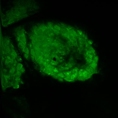
We also did images of a green fluorescent mouse (a mouse full of green fluorescent protein). This is the ear, at the surface:

All the round things are cells. The picture has black gaps where there's no tissue in this plane (this method takes a very, very thin optical "slice" and nothing else).

The idea is very cool because no one can take pictures that deep into tissue without injecting a dye or something. We're supposed to be seeing just hemoglobin (no melanin, this is a nude mouse). You can see blood vessels in the picture, they're the white squiggly things of various sizes (capillaries are very squiggly). However, they're not very bright / clear / pretty. Also, there's a bunch of crap in the image: all those speckled black stuff in the upper half. We're not sure where it's coming from.
We also did images of a green fluorescent mouse (a mouse full of green fluorescent protein). This is the ear, at the surface:

All the round things are cells. The picture has black gaps where there's no tissue in this plane (this method takes a very, very thin optical "slice" and nothing else).