Striptease

Tell me what you see
It sometimes takes extraordinary effort for doctors to test patients. In this respect, ophthalmologists have one of the hardest jobs. In fact, in the Ophtha clinic, the whole consult is 90% percent testing, and 10% interview.
As with any consult, a physician starts with the patient's general information, his chief complaint, and a rundown of the present illness. A survey of systems is done (does the patient have other related/unrelated symptoms?) and then Past, Family and Personal/Social histories are then elicited, focusing on ophthalmologic problems. All of this is done is a span of 5 to 10 minutes. You'd be surprised how fast you get once you get the hang of it (students can spend as much as 30 minutes on a patient!!!).
Then comes the fun part, which is the 5 point examination.
The first test is the reading of the Snellen chart., like the one above This assesses distance vision, visual acuity, and can also pick up errors of refraction. (Click on the middle picture to perform a customized visual acuity test)


A gross examination is then done in order to assess any visible defects in the orbital and circumorbital anatomy.


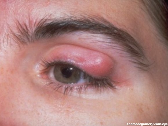
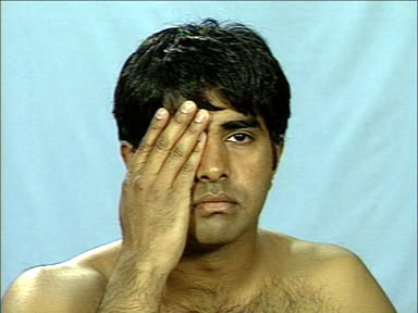
Once this is done, eye movements are assessed in order to gauge extraocular muscle dysfunctions. The patient is supposed to follow the examiner's finger in an H configuration using his eyes and without moving his head. Each eye is tested alone, followed by "conjugate" testing, where both eyes are made to follow the examiner's finger. Some defects are illustrated below.


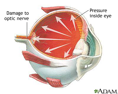
Tonometry, the next test, tests for intraocular pressure. Without the use of instruments, the findings could be reported as hard (as your forehead), firm (as the tip of your nose), and soft (as your lips). The Sentro Oftalmologico in PGH, however, is fully equipped with instruments, including a variety of tonometers. Tonometry is important in assessing for glaucoma. If there is an suspicion, however, of eye puncture, this test (at least the manual one) should be avoided.

Finally, the patient undergoes an internal eye exam, or the fundoscopy. The difficulty of the fundoscopy lies in the fact that we are looking through the pupil, which is normally only 3-4 mm large when exposed to light. Medications may also used to dilate the pupil.


The full ophthalmologic examination is available on video here.
Now, imagine doing that to at least 20 patients a day...
But I hope that allays some fears people have during an ophthalmologic consult.