Diagnostic Imaging II Study Review: Arthridities
Which condition demonstrates nonuniform joint space narrowing, osteophytes, subchondral sclerosis, & subchondral cysts?
--DJD aka degenerative joint disease, I have it in my left pinkie DIP dt tabbing on computers

Which condition presents with triangular sclerosis only at the iliac portion of the lower SI joint?
--osteitis condensans ilii
--mc in females after carrying a fetus to term
--chronic low back pain, SI mobility mb incr or decreased
--completely regresses on its own

Is osteitis condensans ilii more commonly unilateral or bilat?
--bilat & symmetric sclerosis (text says asymmetric (364))
Is osteitis condensans ilii more commonly found in males or females?
--females, childbearing age
Osteitis pubis is commonly associated with which medical procedure?
--surgery near the pubic symphysis
--operations on lower urinary tract esp suprapubic or retropubic prostatectomies
--widening and sclerosis of the symphysis pubis

What is the difference b/w marginal & non-marginal syndesmophytes?
--syndesmophyte = an osseous excrescence attached to a ligament
--non-marginal syndesmophytes don’t come from corners, found in psoriatic & reiters, tends to skip levels in this dzs

--marginal more in ankylosing spondylitis, shiny corners-->bamboo spine

What systemic condition is commonly found in pts w/ diffuse idiopathic skeletal hyperostosis (DISH)?
--diabetes, also dyslipidemia and hyperuricemia

--flowing calcifications and ossifications along the anterolateral aspect of at least 4 contiguous vertebral bodies, with or without osteophytes
--preservation of disk height in the involved areas and an absence of excessive disk disease
--absence of bony ankylosis of facet joints and absence of sacroiliac erosion, sclerosis, or bony fusion, although narrowing and sclerosis of facet joints are acceptable


Dysphagia is common in which arthritic condition & why?
--DISH dt spinal involovment, esp calcification of the anterior longitudinal ligament & extraspinal ligaments may impinge on esophageal space
--this condition has relative maintenance of the disc height and absence of posterior joint fusion or sacroiliitis; there is also ossification of the PLL
--dish present in 12% of people over 40!!!
What part of the spine is DISH most commonly found?
--thorax, lower cervicals, upper lumbar, SI
List the radiographic findings of neurotrophic arthropathy:
--joint changes secondary to overuse/abuse secondary to loss of sensation
--6 D’s: distension, density increase, debris, dislocation, disorganized, destruction
--there is also the abbreviated 3Ds dislocation, destruction & degeneration
--CHARCOT JOINTS = destroyed

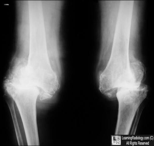


Which conditions may result in neurotrophic arthropathy:
--impaired pain perceptions or proprioception with repeated unrecognized trauma
--DM, alcoholism, tabes dorsalis (dt syphillis), paralysis, syringomyelia
What is synoviochondrometaplasia?

--benign disorder marked by metaplasia of hyperplastic synovium to hyaline cartilage
--hyaline cartilage calcifies and detaches from the synovium to form loose bodies (up to 3 cm in diameter) within the joint, tendon sheath, or bursa
--involves large wt-bearing joints
--most common in knee; occasionally bilateral
--never in the spine
--see: multiple radiodense loose bodies in the joint capsule, tendon sheath or bursa
--pressure erosions, widened joint space, and secondary degeneration
--Sx: intermittent pain, joint swelling, stiffness, “joint locking” episodes
Name the commone sites of involvment of RA in the hand & wrist:
--HANDS: MCP’s & PIP’s; marginal erosions (irregular, no sceroltic margin)

--radial margins of 2nd & 3rd MC head

--Boutonniere (DIP extend, PIP flex)

--Swan neck deformities (DIP flex, PIP extend)

--Ulnar deviation at MCP joint

--WRISTS: ulnar styloid erosion
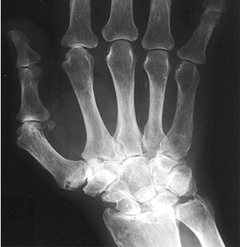
--uniform loss of radiocarpal joint
--erosions of triquetrum-pisiform
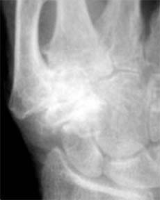
--spotty carpal sign
--pancarpal involvement
--scapholunate dissociation
What is a marginal erosion and what categoy of arthritis isit seen with?
--RA, in the radial side of the metacarpals
What is the significance of widening of the atlanto-dental interspace?
--enlarged; N <3mm in adult and <5 in child
--mbdt rupture of TV ligament-->instability of C-1 and C-0
--can lead to direct compression of the brainstem or excessive kyphosis
--odontoid/dens erosions
--atlanto-axial subluxations-->pseudobasilar invagination
--facet involvement; stair-stepping (anterior-lysthesis; can get with DJD as well)
--IVD involvement
Which conditions demonstrate laxity of the transverse ligament?
--RA, DJD
--SLE, Down’s syndrome
Is SI involvment common in RA?
--No, usu C-spine
--if involved usu minimal sclerosis; uni or bilat & asymmetric
Describe the radiographic difference b/w RA & Psoriatic Arthritis in the hand & wrist.
--PSORIATIC: DIPS, assymetrial, similar to RA w/o hyperemia, soft tissue swelling, no osteopenia, yes erosions, fluffy periostitis, narrowed or widened joint space, osseous fusion, acro-osteolysis, pencil in cup deformity, ray patter (all 3 jts in 1 digit)

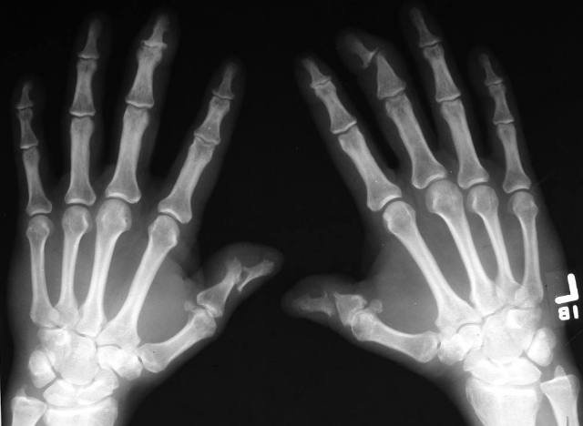
--Psoriatic in SI: bilateral, asymmetric, unilateral, indistinct margins
--Psoriatic in spine: nonmarginal syndesmophyes, thicker growths, assymetrical, coarse, esp at TL juct, atlantoaxial instability, less facet involvemnt
--RA in hands & wrists: PIPs, uniform, bilateral, MCP’s (Haygarth’s nodes), marginal erosions (irregular, no sceroltic margin, Rat bite lesion), radial margins of 2nd & 3rd MC head, Boutonniere (DIP extend, PIP flex), Swan neck deformities (DIP flex, PIP extend), Lanois deformity, ulnar deviation at MCP joint, bilateral and symmetrical and uniform joint space loss, most common Protusio Acetabuli in pelvis
What is the gender incidence of rhemuatoid arthritis?
--F:M 3:1 until 40, then 1:1
What is the first site of involvement with ankylosing spondylitis?
--SI, lower 2/3 (synovial portion) of the joint
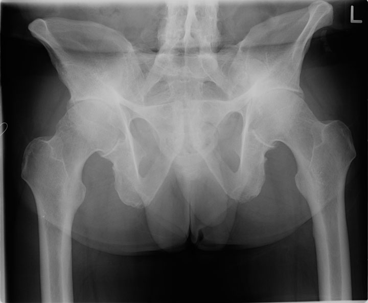
What is the second site of involvemenet with AS?
--spine, usu at thoracolumbar junction, sometimes lumbosacra junction

Is the SI involvement usually unilateral or bilateral in AS?
--bilateral

What is the gender incidence of the vertebral column & pelvis in ankylosing spondylitis?
--male 9:1
--onset at 20-60 yo (peak 40-50 yo)
What condition demonstrates squaring of the vertebral body?
--AS

What is the shiny corner sx?
--increased radiodensity of the corners of the vertebral body related to osteitis
--reactive sclerosis that is seen in AS
--bone sclerosis at the anterior vertebral margins associated with Romanus lesions in patients who have AS
--Romanus lesion is found at the insertion of the outer annulus fibrosus into the anterior corners of the vertebral bodies
What is a “carrot stick” fracture?
--fracture of an ankylosed segment of vertebrae in AS
--usu causes paralysis
Which condition demonstrates similar SI joint & vertebral column findings to AS?
--enteropathic arthropathy
--secondary to: Ulcerative colitis, Crohn’s disease, Whipple’s disease, Salmonella, Shigella, Yersinia
Which two seronegative sondyloarthropathies demonastrate non-marginal syndesmophytes and peipheral arthritis?
--psoriatic and Reiters
--non-marginal syndesmophytes-thicker, not throughout the spine like AS
Reversible deformities of the hand are seen in which condition?
--Systemic Lupus Erythematous (SLE)
--ulnar deviation, like RA, but pt can overcome w/muscle contraction or pushing hand down onto a table
--ligaments are lax, but jts are not destroyed
What is acro-osteolysis & which condition demonstrates this finding?
--resorbtion of the extremities (ie: distal phalanx "tufts")
--#1: scleroderma, also in psoriatic, SLE, hyperparathyroid
What is the overhanging margin sign & which condition is this seen in?


--pathoneumonic finding in Gout, a C-shaped erosion that sticks out
--sclerotic margin outside the joint capsule


What structures are primarily involved in CPPD (calcium pyrophosphate dihydrate crystal deposition disease)?
--#1 wrist, triangular fibrocartilage distal to ulnar styloid
--#2 knee, meniscus


--#3 pubic symphysis
--involves calcification of cartilage-->chondrocalcinosis in intermediate layer
--fibrous & hyaline cartilage
--fibrous in meniscus and triangle
--hyaline at end of bones: calcification parallel to cortex, thin, linear
--AKA PSEUDOGOUT onset after 30 peak at 60
--Dx: aspiration of synovial fluid
What structures are primarily involved in HADD (hydroxyapatite deposition dz)?
--bursae and tendons, calcification outside joint near insertion
--shoulder (supraspinatus) and hip (glut med tendon)


What are the characteristics of juvenile rheumatoid arthritis??
Still's disease is a systemic, inflammatory disorder that occurs in children (in whom it is called both "Still's disease" and "systemic-onset juvenile idiopathic arthritis") and adults (in whom it is designated as "adult-onset Still's disease"). In both children and adults the clinical picture includes symmetrical arthritis, fever, fatigue, rash, leukocytosis, adenopathy, and hepatosplenomegaly. Tx: prednisone, 20 mg bid==>joint inflammation, fatigue, fever, anemia, leukocytosis, and thrombocytosis should improve rapidly.
--DJD aka degenerative joint disease, I have it in my left pinkie DIP dt tabbing on computers

Which condition presents with triangular sclerosis only at the iliac portion of the lower SI joint?
--osteitis condensans ilii
--mc in females after carrying a fetus to term
--chronic low back pain, SI mobility mb incr or decreased
--completely regresses on its own

Is osteitis condensans ilii more commonly unilateral or bilat?
--bilat & symmetric sclerosis (text says asymmetric (364))
Is osteitis condensans ilii more commonly found in males or females?
--females, childbearing age
Osteitis pubis is commonly associated with which medical procedure?
--surgery near the pubic symphysis
--operations on lower urinary tract esp suprapubic or retropubic prostatectomies
--widening and sclerosis of the symphysis pubis

What is the difference b/w marginal & non-marginal syndesmophytes?
--syndesmophyte = an osseous excrescence attached to a ligament
--non-marginal syndesmophytes don’t come from corners, found in psoriatic & reiters, tends to skip levels in this dzs

--marginal more in ankylosing spondylitis, shiny corners-->bamboo spine

What systemic condition is commonly found in pts w/ diffuse idiopathic skeletal hyperostosis (DISH)?
--diabetes, also dyslipidemia and hyperuricemia

--flowing calcifications and ossifications along the anterolateral aspect of at least 4 contiguous vertebral bodies, with or without osteophytes
--preservation of disk height in the involved areas and an absence of excessive disk disease
--absence of bony ankylosis of facet joints and absence of sacroiliac erosion, sclerosis, or bony fusion, although narrowing and sclerosis of facet joints are acceptable


Dysphagia is common in which arthritic condition & why?
--DISH dt spinal involovment, esp calcification of the anterior longitudinal ligament & extraspinal ligaments may impinge on esophageal space
--this condition has relative maintenance of the disc height and absence of posterior joint fusion or sacroiliitis; there is also ossification of the PLL
--dish present in 12% of people over 40!!!
What part of the spine is DISH most commonly found?
--thorax, lower cervicals, upper lumbar, SI
List the radiographic findings of neurotrophic arthropathy:
--joint changes secondary to overuse/abuse secondary to loss of sensation
--6 D’s: distension, density increase, debris, dislocation, disorganized, destruction
--there is also the abbreviated 3Ds dislocation, destruction & degeneration
--CHARCOT JOINTS = destroyed


Which conditions may result in neurotrophic arthropathy:
--impaired pain perceptions or proprioception with repeated unrecognized trauma
--DM, alcoholism, tabes dorsalis (dt syphillis), paralysis, syringomyelia
What is synoviochondrometaplasia?

--benign disorder marked by metaplasia of hyperplastic synovium to hyaline cartilage
--hyaline cartilage calcifies and detaches from the synovium to form loose bodies (up to 3 cm in diameter) within the joint, tendon sheath, or bursa
--involves large wt-bearing joints
--most common in knee; occasionally bilateral
--never in the spine
--see: multiple radiodense loose bodies in the joint capsule, tendon sheath or bursa
--pressure erosions, widened joint space, and secondary degeneration
--Sx: intermittent pain, joint swelling, stiffness, “joint locking” episodes
Name the commone sites of involvment of RA in the hand & wrist:
--HANDS: MCP’s & PIP’s; marginal erosions (irregular, no sceroltic margin)

--radial margins of 2nd & 3rd MC head

--Boutonniere (DIP extend, PIP flex)

--Swan neck deformities (DIP flex, PIP extend)

--Ulnar deviation at MCP joint
--WRISTS: ulnar styloid erosion

--uniform loss of radiocarpal joint
--erosions of triquetrum-pisiform

--spotty carpal sign
--pancarpal involvement
--scapholunate dissociation
What is a marginal erosion and what categoy of arthritis isit seen with?
--RA, in the radial side of the metacarpals
What is the significance of widening of the atlanto-dental interspace?
--enlarged; N <3mm in adult and <5 in child
--mbdt rupture of TV ligament-->instability of C-1 and C-0
--can lead to direct compression of the brainstem or excessive kyphosis
--odontoid/dens erosions
--atlanto-axial subluxations-->pseudobasilar invagination
--facet involvement; stair-stepping (anterior-lysthesis; can get with DJD as well)
--IVD involvement
Which conditions demonstrate laxity of the transverse ligament?
--RA, DJD
--SLE, Down’s syndrome
Is SI involvment common in RA?
--No, usu C-spine
--if involved usu minimal sclerosis; uni or bilat & asymmetric
Describe the radiographic difference b/w RA & Psoriatic Arthritis in the hand & wrist.
--PSORIATIC: DIPS, assymetrial, similar to RA w/o hyperemia, soft tissue swelling, no osteopenia, yes erosions, fluffy periostitis, narrowed or widened joint space, osseous fusion, acro-osteolysis, pencil in cup deformity, ray patter (all 3 jts in 1 digit)


--Psoriatic in SI: bilateral, asymmetric, unilateral, indistinct margins
--Psoriatic in spine: nonmarginal syndesmophyes, thicker growths, assymetrical, coarse, esp at TL juct, atlantoaxial instability, less facet involvemnt
--RA in hands & wrists: PIPs, uniform, bilateral, MCP’s (Haygarth’s nodes), marginal erosions (irregular, no sceroltic margin, Rat bite lesion), radial margins of 2nd & 3rd MC head, Boutonniere (DIP extend, PIP flex), Swan neck deformities (DIP flex, PIP extend), Lanois deformity, ulnar deviation at MCP joint, bilateral and symmetrical and uniform joint space loss, most common Protusio Acetabuli in pelvis
What is the gender incidence of rhemuatoid arthritis?
--F:M 3:1 until 40, then 1:1
What is the first site of involvement with ankylosing spondylitis?
--SI, lower 2/3 (synovial portion) of the joint

What is the second site of involvemenet with AS?
--spine, usu at thoracolumbar junction, sometimes lumbosacra junction

Is the SI involvement usually unilateral or bilateral in AS?
--bilateral

What is the gender incidence of the vertebral column & pelvis in ankylosing spondylitis?
--male 9:1
--onset at 20-60 yo (peak 40-50 yo)
What condition demonstrates squaring of the vertebral body?
--AS

What is the shiny corner sx?
--increased radiodensity of the corners of the vertebral body related to osteitis
--reactive sclerosis that is seen in AS
--bone sclerosis at the anterior vertebral margins associated with Romanus lesions in patients who have AS
--Romanus lesion is found at the insertion of the outer annulus fibrosus into the anterior corners of the vertebral bodies
What is a “carrot stick” fracture?
--fracture of an ankylosed segment of vertebrae in AS
--usu causes paralysis
Which condition demonstrates similar SI joint & vertebral column findings to AS?
--enteropathic arthropathy
--secondary to: Ulcerative colitis, Crohn’s disease, Whipple’s disease, Salmonella, Shigella, Yersinia
Which two seronegative sondyloarthropathies demonastrate non-marginal syndesmophytes and peipheral arthritis?
--psoriatic and Reiters
--non-marginal syndesmophytes-thicker, not throughout the spine like AS
Reversible deformities of the hand are seen in which condition?
--Systemic Lupus Erythematous (SLE)
--ulnar deviation, like RA, but pt can overcome w/muscle contraction or pushing hand down onto a table
--ligaments are lax, but jts are not destroyed
What is acro-osteolysis & which condition demonstrates this finding?
--resorbtion of the extremities (ie: distal phalanx "tufts")
--#1: scleroderma, also in psoriatic, SLE, hyperparathyroid
What is the overhanging margin sign & which condition is this seen in?


--pathoneumonic finding in Gout, a C-shaped erosion that sticks out
--sclerotic margin outside the joint capsule


What structures are primarily involved in CPPD (calcium pyrophosphate dihydrate crystal deposition disease)?
--#1 wrist, triangular fibrocartilage distal to ulnar styloid
--#2 knee, meniscus


--#3 pubic symphysis
--involves calcification of cartilage-->chondrocalcinosis in intermediate layer
--fibrous & hyaline cartilage
--fibrous in meniscus and triangle
--hyaline at end of bones: calcification parallel to cortex, thin, linear
--AKA PSEUDOGOUT onset after 30 peak at 60
--Dx: aspiration of synovial fluid
What structures are primarily involved in HADD (hydroxyapatite deposition dz)?
--bursae and tendons, calcification outside joint near insertion
--shoulder (supraspinatus) and hip (glut med tendon)


What are the characteristics of juvenile rheumatoid arthritis??
Still's disease is a systemic, inflammatory disorder that occurs in children (in whom it is called both "Still's disease" and "systemic-onset juvenile idiopathic arthritis") and adults (in whom it is designated as "adult-onset Still's disease"). In both children and adults the clinical picture includes symmetrical arthritis, fever, fatigue, rash, leukocytosis, adenopathy, and hepatosplenomegaly. Tx: prednisone, 20 mg bid==>joint inflammation, fatigue, fever, anemia, leukocytosis, and thrombocytosis should improve rapidly.