if you blink you might miss it...
I've decided to start writing in my LJ again. We all know this is a fad that never lasts with me but I was feeling kind of guilty reading Tsadida's monthly updates that usually list me as 'still in grad school... we think' lol. Speaking of which, thanks for the Christmas cards peoples. The cards I bought to write last year are still on my dresser. If I ever send them it will be horribly off season and a bit of a surprise :x
I was going through all of my data for an upcoming committee meeting (the yearly 'are you on schedule to graduate soon' type deal) and I decided to post some of my data. I can only publish the wild type result really (can't post mutant data until it's at least presented at a meeting).
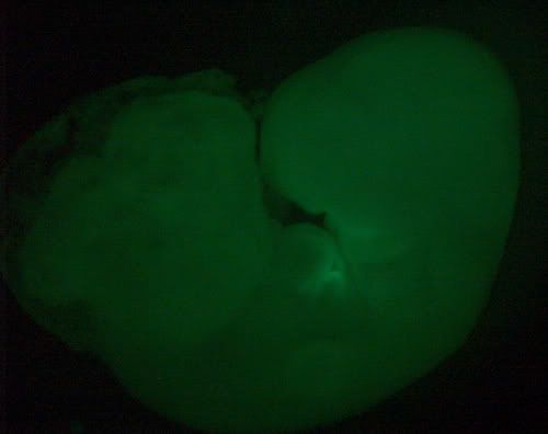
This is a picture of a mouse that I grew in culture for 28 hours. Basically once the embryos are taken from the mom you have to be really careful to keep the placenta and yolk sac (the thing that has all the blood vessels leading to the placenta) intact. You grow them up in a blood serum replacement in little glass bottles that are on this ferris wheel type contraption to keep temperature and oxygen levels regulated. It's a rather tricky procedure and I was really excited when I managed to get it working. This embryo has been surface labeled with CMFDA (a green dye). I'm kind of asking a geography question with this project and if I ever get the cell death label to work it will be cool to look at ._.;;
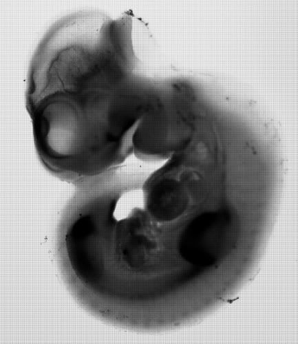
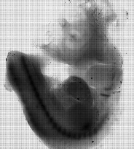
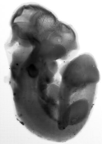
Another annoying thing that I do in the lab (annoying as in really easy to screw up) is in situ hybridization. Basically you make a molecular probe which identifies and hybrizes with (grabs on to) RNA in a cell. Anywhere you see dark stuff (blue if these pictures were in color) the RNA you are looking at is present in the embryo. These were my first attempts at probe making. I made probes for Bmp4, Pax1, and Tbx1.
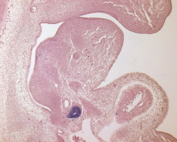
Recently I have been doing a lot of in situ hybridization. However instead of doing wholemount (whole embryo) in situ like those previous pictures, I have been doing section in situ. It's a bit cleaner and I've got it down to a science (yes I know that's a bad joke). This first picture is a saggital section from an E11.5 wild type embryo (11.5 is close to the start of thymus development and pretty much in the middle of mouse development - they're born at 18 days or so). The purple stuff is just normal tissue, I've used a hemotoxylin and eosin stain (hemotoxylin dyes the nucleus blue and eosin dyes the cytoplasm red making the whole thing purple). The blue thing is a probe for Foxn1 and it labels the thymus. The big gap to the left of the thymus is trachea and the roundish cavity to the right of the thymus is heart.
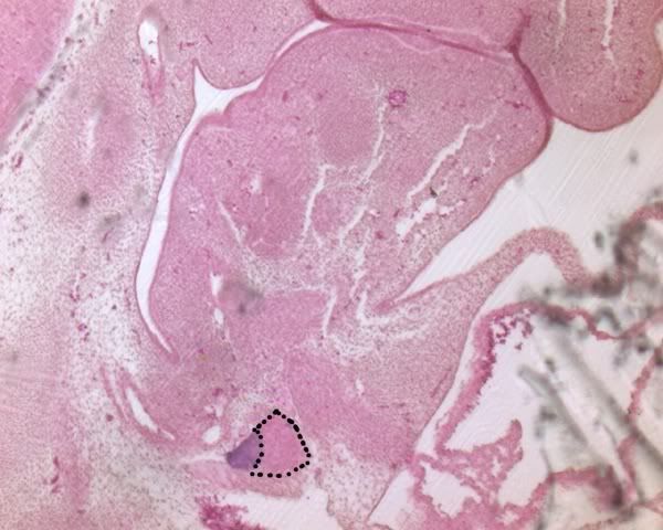
The other half of the third pharyngeal pouch (that place where the thymus comes from) forms the parathyroid. You can detect parathyroid using a marker for Gcm2. This is a picture of an E12.5 embryo. You can see the Gcm2 (parathyroid) domain in blue. I have outlined the thymus domain. Once again trachea is to the left and heart is to the right.
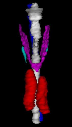
One other thing I do a bit of in the lab is 3-D reconstruction. Basically once I do an experiment on an embryo, I section it and take a picture of every section in the series. Then I use a computer program to trace the tissues I'm interested in and make a 3-D model. Here's one that shows parathyroid (aqua) thymus (red) thyroid (purple) and trachea (white) at e14.5 (but it looks the same as it will at an adult stage).
That's a pretty good summary of the types of stuff I do in lab. Everything else is molecular work (DNA extraction, probe making, genotyping). I'm trying to perfect the finer are of antibody staining at the moment (it's really pretty stuff) and if I ever get it to work, I'll post some pretty pictures (cross your fingers). I'll be posting more later, honest <.<; probably something about crazy adventures in gaming.
I was going through all of my data for an upcoming committee meeting (the yearly 'are you on schedule to graduate soon' type deal) and I decided to post some of my data. I can only publish the wild type result really (can't post mutant data until it's at least presented at a meeting).

This is a picture of a mouse that I grew in culture for 28 hours. Basically once the embryos are taken from the mom you have to be really careful to keep the placenta and yolk sac (the thing that has all the blood vessels leading to the placenta) intact. You grow them up in a blood serum replacement in little glass bottles that are on this ferris wheel type contraption to keep temperature and oxygen levels regulated. It's a rather tricky procedure and I was really excited when I managed to get it working. This embryo has been surface labeled with CMFDA (a green dye). I'm kind of asking a geography question with this project and if I ever get the cell death label to work it will be cool to look at ._.;;


Another annoying thing that I do in the lab (annoying as in really easy to screw up) is in situ hybridization. Basically you make a molecular probe which identifies and hybrizes with (grabs on to) RNA in a cell. Anywhere you see dark stuff (blue if these pictures were in color) the RNA you are looking at is present in the embryo. These were my first attempts at probe making. I made probes for Bmp4, Pax1, and Tbx1.

Recently I have been doing a lot of in situ hybridization. However instead of doing wholemount (whole embryo) in situ like those previous pictures, I have been doing section in situ. It's a bit cleaner and I've got it down to a science (yes I know that's a bad joke). This first picture is a saggital section from an E11.5 wild type embryo (11.5 is close to the start of thymus development and pretty much in the middle of mouse development - they're born at 18 days or so). The purple stuff is just normal tissue, I've used a hemotoxylin and eosin stain (hemotoxylin dyes the nucleus blue and eosin dyes the cytoplasm red making the whole thing purple). The blue thing is a probe for Foxn1 and it labels the thymus. The big gap to the left of the thymus is trachea and the roundish cavity to the right of the thymus is heart.

The other half of the third pharyngeal pouch (that place where the thymus comes from) forms the parathyroid. You can detect parathyroid using a marker for Gcm2. This is a picture of an E12.5 embryo. You can see the Gcm2 (parathyroid) domain in blue. I have outlined the thymus domain. Once again trachea is to the left and heart is to the right.

One other thing I do a bit of in the lab is 3-D reconstruction. Basically once I do an experiment on an embryo, I section it and take a picture of every section in the series. Then I use a computer program to trace the tissues I'm interested in and make a 3-D model. Here's one that shows parathyroid (aqua) thymus (red) thyroid (purple) and trachea (white) at e14.5 (but it looks the same as it will at an adult stage).
That's a pretty good summary of the types of stuff I do in lab. Everything else is molecular work (DNA extraction, probe making, genotyping). I'm trying to perfect the finer are of antibody staining at the moment (it's really pretty stuff) and if I ever get it to work, I'll post some pretty pictures (cross your fingers). I'll be posting more later, honest <.<; probably something about crazy adventures in gaming.